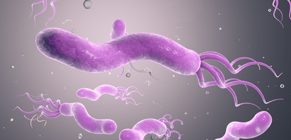
The bacterium Helicobacter pylori, responsible for gastric problems, may also contribute to the severity of bronchiectasis. The study, “Is There Any Role Of Helicobacter pylori In The Pathogenesis Of Non-Cystic Fibrosis Bronchiectasis?” was recently published in the Pediatrics International journal.
Bronchiectasis is a chronic pulmonary disease characterized by inflammation and irreversible bronchi dilatation. The main causes for bronchiectasis in developing countries are infections by three pathogens: Bordetella pertussis, measles, and Mycobacterium tuberculosis. Comparatively, in developed countries the most common causes are immunological disorders and cystic fibrosis (CF). Bronchiectasis incidence in children, despite low, is usually severe enough to lead to significant morbidity and mortality.
Researchers investigated whether Helicobacter pylori, a gram-negative bacterium causing gastric problems (from chronic active gastritis to gastric carcinoma) may also cause non-CF bronchiectasis. Previous reports detected Helicobacter pylori in the trachea-bronchial aspirates of mechanically ventilated patients . Researchers also investigated the bacterium role in gastroesophageal reflux.
The team enrolled 41 bronchiectasis patients and 16 controls ages 5 to 18 at the Hacettepe University School of Medicine Department of Pediatric Pulmonology in Ankara, Turkey. Helicobacter pylori presence was determined in both bronchiectasis patients and controls by polymerase chain reaction, culture in gastric juice, and bronchoalveolar lavage fluid, as well as with a urea breath test.
Gastroesophageal reflux was detected by monitoring pH in a 24-hour period or by a specific technique called scintigraphy, a noninvasive imaging technique where patients are administered with radionuclides, or radioactive elements called isotopes or tracers which are usually attached to drugs and travel to a specific organ in the body that is then analyzed. The retrieved information was then correlated with the severity and extent of bronchiectasis, measured by computerized tomography scoring.
Researchers found nine patients (22 percent) in the bronchiectasis group and three patients (18.8 percent) in the control group with bronchoalveolar lavage fluid positive for Helicobacter pylori. In the gastric juice, 16 bronchiectasis patients (39 percent) and seven controls (43.8 percent) were positive for the bacterium, while the urea breath test gave positive results in 11 patients (26.8 percent) and three controls (18.8 percent), respectively.
There was no significant difference between the bronchiectasis group and the control group for any of the tested samples, including bronchoalveolar lavage fluid, gastric juice, and the urea breath test, suggesting that Helicobacter pylori has no role in the pathogenesis of bronchiectasis.
Notably, however, researchers found that computerized tomography scoring was higher in bronchiectasis patients with bronchoalveolar lavage fluid positive for Helicobacter pylori when compared to patients whose samples were negative. Hence, the authors believe that Helicobacter pylori may contribute to the severity of the disease.

