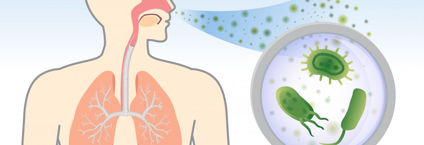
An abundance of Pseudomonas aeruginosa bacteria in the airways of patients with non-cystic fibrosis (CF) bronchiectasis is linked to greater inflammation by dampening expression of a gene called PPARγ, a study reports.
The study, “PPARγ is reduced in the airways of non-CF bronchiectasis subjects and is inversely correlated with the presence of Pseudomonas aeruginosa,” was published in the journal PLOS ONE. Its scientists also suggested that available treatments for diabetes that work as PPARy agonists may be of help to these patients.
Patients with non-CF bronchiectasis with a lung infection caused by P. aeruginosa bacteria tend to have poorer outcomes, experiencing faster lung function decline. But the reason why has been unclear.
A protein called peroxisome proliferator-activated receptor gamma (PPARγ) is known to exert anti-inflammatory effects, and to be present in lower than normal levels in the airways of CF patients, particularly those infected with P. aeruginosa.
The PPARγ protein — which is produced based on genetic information on the PPARy gene — senses specific fat, or lipid, molecules inside cells, shutting down or activating certain genes controlling inflammation and metabolism.
Unlike in CF patients, the potential role of the PPARγ gene in patients with non-CF bronchiectasis has not been investigated.
Researchers in Australia examined the expression of PPARγ in airway cells of people with non-CF bronchiectasis. A total of 35 patients and 20 healthy controls were included in the study. All were participants in the Phase 2a trial BLESS (ACTRN12609000578202), and none had been treated with macrolide antibiotics.
Researchers also analyzed the relationship between PPARγ expression, the number of immune cells, and the presence of important infectious agents, including P. aeruginosa, in the lungs.
Results showed that PPARγ was significantly less expressed in the airways of non-CF bronchiectasis patients, with levels nearly half those observed in healthy controls.
Importantly, the low expression levels were significantly associated with the presence of high numbers of P. aeruginosa in mucus (sputum) of patients.
No significant association was seen between PPARγ expression and total bacterial levels or levels of Haemophilus influenzae.
Regarding immune cells in the airway fluid, higher levels of PPARγ expression were weakly but significantly associated with increased numbers of macrophages, key immune cells for inflammation. No other immune cells analyzed showed a significant link with PPARγ gene expression, suggesting that “macrophages may be the primary source of PPARγ in non-CF bronchiectasis airways,” the researchers wrote.
In agreement with observations made in CF patients, these data demonstrate that “PPARγ is expressed in low levels in the airways of non-CF bronchiectasis subjects, despite an aggressive inflammatory response. This low level PPARγ expression is particularly associated with the presence of high levels of P. aeruginosa, and may represent an intrinsic link with this bacterial pathogen.”
While further work is necessary, the study “supports the notion that low levels of traditional anti-inflammatory molecules may also play a role in disease due to the inability of the host to rein in exuberant inflammation,” the researchers wrote.
Of note, “PPARγ agonists [activators] are commercially available treatments for Type 2 Diabetes Mellitus and may offer a potential therapeutic agent to aid in the treatment of inflammatory airways diseases” like bronchiectasis, they suggested.

